
Complex Orthopaedics
Orthopaedics is a surgical speciality that treats bone and joint issues in dogs and cats. It can be extremely complex, necessitating extensive training and experience to execute correctly.
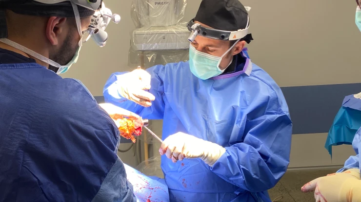
At my veterinary clinic, I perform complex orthopaedic procedures and am committed to providing our patients with the best possible care. Through advanced surgical techniques, I’m able to provide relief for pets who are suffering from a wide range of orthopaedic issues such as fractures, deformities of legs and degenerative joint diseases such as cruciate ligament disease.
Types of Complex Orthopaedic Surgeries
Tibial plateau leveling osteotomy (TPLO)
What is a TPLO?
Tibial plateau leveling osteotomy (TPLO) is the most common surgeries I do. It is used to treat dogs with cranial cruciate ligament tears. To date, I have performed over 3000 TPLO procedures and success rates are above 99%. TPLO was invented and patented by Dr Barclay Slocum (USA) in 1993 and has since been refined and modernised to the procedure it is today. The recovery is quick with 1 night in hospital and 6-8 weeks recovering.
Fracture Stabilisation
What are bone fractures?
How are fractures treated?
Fractured bones need to be stabilised and supported until they can handle the body’s weight and movement without assistance. The most common surgical procedures used to treat fractures are bone plates or minimally invasive osteosynthesis.
Bone Plates
What are bone plates?
Bone plates are specialised fixation devices used to treat fractures by holding the broken bone fragments in place while they’re healing. They are generally left in place once the bone has healed.
Non-locking plates work by compressing against the bone which helps to keep them in place and reduce the risk of loosening or movement. Screws are attached to the bone to hold the plate in place.
Locking plates are different from non-locking plates in that they have screws that help to hold the bone fragments in place. The advantage of locking plates is that they don’t rely on compression between the bone and the plate to keep them in place. Because of this, the plate doesn’t have to be shaped perfectly to fit the bone, they are easier to put in place, less time is needed in surgery and they create a stronger fixation.
Minimally invasive osteosynthesis
Minimally invasive osteosynthesis (MIO) is a type of fracture repair that uses fluoroscopy to guide the placement of screws, pins or plates in order to fix broken bones.
Fluoroscopy is a medical imaging technique that creates a moving X-ray image, like an X-ray movie. During fluoroscopy surgery, an X-ray beam moves through the body and the resulting image is transmitted to a monitor. This allows the surgeon to see inside the body and helps guide the placement of plates and screws during surgery.
The advantage of MIO is that it allows for a quicker and less invasive surgery, with shorter recovery times and fewer complications. It is also extremely precise, meaning that the screws are placed in exactly the right place to ensure ideal healing of the fracture.

What are angular and antebrachial limb deformities?
It can lead to:
- shortened limbs
- bowing of bones
- partial displacement of joints
- bending or twisting of growing bone
Causes and symptoms of angular and antebrachial limb deformities
Some of the common symptoms of angular limb deformity include:
- limping or lameness
- swelling and pain
- unusual posture, gait or movement
How is angular limb deformity diagnosed?
A physical examination and X-rays are used to diagnose angular limb deformities. During a physical exam, the patient’s way of walking and range of motion is observed. The joints near the deformity are felt for signs of pain and swelling.
Since every deformity is different, the diagnosis of angular limb deformity is typically made through several imaging techniques. Diagnosis starts with an X-ray to determine the extent of the deformity and evaluate any areas of concurrent joint damage. The advantage of X-rays is that they are quick, non-invasive and effective for simple abnormalities.
However, X-rays are limiting in that they cannot provide a 3D picture of the bone and can miss more complex deformities. In cases where more detailed imaging is needed, a computer tomography (CT) scan is used to create a 3D model of the bone.
Creating 3D models allows us to visualise and control the limb in three dimensions and plan surgeries. The 3D model can be used to create what is called a ‘surgical guide’. The surgical guide helps us position plates and screws in the correct location and ensure the bone is repaired in the correct position and orientation.
We can take the process even further by 3D printing of a physical replica of the deformed limb and surgical guides. This allows us to create highly accurate replicas that are tailored precisely to each individual patient’s needs.
What are the treatment options for angular limb deformity?

Complex Spinal Surgeries
What is a spinal fracture?
Spinal fractures are breaks in one or more of the vertebrae, which are the bones that make up the spinal column.
Fractures can be complete (when the vertebrae breaks into two pieces) or incomplete (where the vertebrae isn’t completely broken).
Spinal fractures can be dangerous because the vertebrae surround the spinal cord so damage to the vertebrae can cause serious nerve damage and paralysis.
How are spinal fractures diagnosed?
Initially, animals are examined for consistent symptoms of spinal fracture including:
- significant pain when the spine is touched
- reduced free movement in the limbs and/or tail
- fewer or no reflexes
- decreased sensation
- decreased bladder and anal tone
A definitive diagnosis is done by X-ray for visual evidence of vertebrae fracture. A CT scan can also be used to get a more detailed view of damage to the vertebrae, which may not be seen in an X-ray. MRI scanning is used to assess spinal cord damage which is not as easily seen on a CT scan. CT scanners are good for bones/joints, whereas MRIs are better for ‘soft tissues’ such as spinal cord and nerves.
How are spinal fractures treated?
In some rare cases, non-surgical treatment options may be available. These can heal with pain medication and strict limitation of movement.
Unstable spinal fractures are managed with surgery. The aim is to stabilise and re-align the fractured vertebrae. This is done by implanting screws or pins into both sides of the fracture and securing them in place with either a plate or cement.
How long is the recovery?

Intervertebral Disc Disease (IVDD)
What is intervertebral disc disease?
Intervertebral disc disease is a disease of the spinal cord.
The spinal cord is made up of individual nerve cells that transfer messages between the brain and the rest of the body. It’s surrounded by bones named vertebrae that act as protective casing. In between each of the vertebrae is an intervertebral disk which is fibrocartilaginous (jelly-like). The intervertebral disks provide support, absorb shock and allow the spine to move.
IVDD occurs when the intervertebral discs degrade and become less shock-absorbent. This eventually leads to disc herniation (slipping) and compression of the spinal cord.
Types of intervertebral disc disease (IVDD)
Hansen type I disc disease is most common in small dog breeds and is more likely to occur in dogs over the age of two. In this disease, the inner part of the intervertebral disc becomes herniated (the jelly-like centre sticks out). It’s very painful, can cause paralysis and in the most severe cases requires emergency surgery. Often symptoms occur rapidly.
Hansen type-III disc disease has a sudden onset. It’s usually caused by extensive exercise or trauma which causes the nucleus (inner part of the disc) to tear through the annulus (outer part of the disc). The injury to the spinal cord does not continue to put pressure on the spinal cord and with rehabilitation and physiotherapy, most animals get better without surgery.
What are the symptoms of intervertebral disc disease?
Symptoms of intervertebral disc disease include:
- spinal pain localised to the back or neck
- abnormal posture
- panting and shivering
- unwillingness to move
- loss of sensation
- inability to empty bladder
- progressive weakness or paralysis
How is intervertebral disc disease diagnosed?
At the onset, intervertebral disc disease can be determined by observing an animal’s symptoms. However, advanced imaging is required for a definitive diagnosis.
Spinal radiographs (X-rays of the spine) can be used to look for characteristic changes seen in IVDD such as calcification (hardening) of the intervertebral discs. The problem with this imaging is that it doesn’t give accurate localisation which is needed for surgery.
MRI and CT scans can provide a more accurate diagnosis as they can localise the affected area of the spine and help to provide a better understanding of which vertebrae are involved. They can also be used to assess how much damage has been done to the nerves and spinal cord.
How is intervertebral disc disease treated?
There are different ways to treat IVDD. The first way is called ‘conservative management’. This means that you will take care of your dog at home and give it medicine. It usually involves lots of rest and pain medication to help ease the symptoms. Crate rest is also recommended for at least 4-6 weeks.
If a dog has paralysis due to IVDD, they may require decompressive surgery. This is a procedure that involves opening the spinal cord compartments and removing any herniated material that is compressing the spinal cord. Decompressive surgery is typically only performed in cases of paresis or paralysis where the dog is not able to walk or stand.
Recovery after surgery
Most dogs will require a period of rest and rehabilitation after surgery for IVDD. During this time, the dog should be kept in a crate or small area to protect them from further injury. They should also be provided with plenty of soft bedding to rest.
Anti-inflammatory medication may be prescribed to help reduce inflammation and pain. Physical therapy may also be recommended to help the dog regain strength and mobility. Most dogs will make a full recovery with appropriate treatment, but some may experience long-term problems such as weakness, paralysis or incontinence.

Recovery after surgery
What is lumbosacral disease?
Lumbosacral disease is a condition that affects the lumbosacral spine or the lower back. This area is made up of the lumbar vertebrae and the sacrum (where the lumbar spine meets the pelvis). This area is responsible for supporting the weight of the body. Lumbosacral disease is brought on by the narrowing of the spinal canal, resulting in pressure being placed upon nerves that exit the spine.
What are the symptoms of lumbosacral disease?
Symptoms of lumbosacral disease include:
- lower back pain
- hind limb pain (e.g. limping)
- hind limb pain, lameness and weakness
- hind limb incoordination
- urinary or faecal incontinence
- progressive weakness or paralysis
- progressive weakness or paralysis
How is lumbosacral disease diagnosed?
How is lumbosacral disease treated?
Treatment options include:
- non-surgical management
- decompressive surgery (laminectomy)
- lumbosacral distraction stabilisation surgery
